General physiology
The inner ear contains the sensory organs responsible for hearing and balance. The cochlea is a snail-shaped, bony structure that propagates sound in the inner ear, while the vestibule or labyrinth contributes to balance.
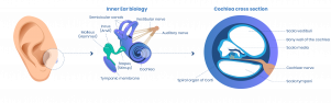
The cochlea
The cochlea is filled with two fluids (endolymph and perilymph), inside the cochlea is the sensory receptor — the Organ of Corti — which contains sensory cells with hair-like structures (hair cells) that are the nerve receptors for hearing.
The bones of the middle ear transform sound into a force that pushes on a membrane (the oval window) in the cochlea, moving the fluid in the cochlea, stimulating the sensory hair cells. Signals from hair cells are transmitted via the auditory nerve as neural impulses to the mid-brain, or the cochlear nucleus, and from there to the hearing region of the brain (auditory cortex).
The vestibule
The vestibule uses the same kind of fluids and detection cells (hair cells) as the cochlea, but to detect motion and orientation. The type of motion detected by a hair cell depends on the structures associated with it (e.g. the semicircular canal or the crystalline otolith of the saccule and utricle). The brain receives, interprets, and processes the signals to create the sensation of balance.